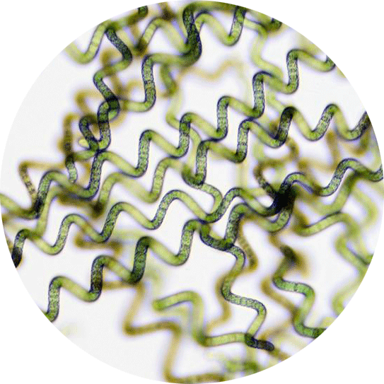
Spirulina under the microscope, a very close and unusual look
In this short article, we thought to share with you a little about the spirulina under the microscope view. You will have a very close encounter. This is very fun and interesting to observe.
(Observation under the microscope is done every week for quality assurance)

Starting with scaling, this will give you the sizing proportions for your own perspective
Right below you can view a result of one of the very first experiments. This is freshly harvested spirulina less than a minute after it was removed/harvested from the water (cultivation medium).
The image demonstrates eyes level view as if you hold spirulina in the palm of your hand.
Spirulina under the microscope
Spirulina at 10x magnification – “Still can’t see much”
In the picture below, while everything still seems to be quite blurry if you look carefully you can certainly start seeing individual elements.
Spirulina at 60x magnification – “Things start to clear”
This picture below shows clear spirulina filaments (i.e. long-chain*). Those spirulina individual filaments are suspended within its growth media.
*Clear explanation and understanding of spirulina filaments is coming.
Spirulina at 200x magnification
In this next image, you can view very clear individual spirulina filaments. As a rule of thumb, if a filament has 4 or more spirals in it, it is a healthy filament. Shorter filaments are not “sick” but the overall health of the culture can be determined through this observation.
You will also notice at the bottom of the picture long straight filaments. This is spirulina’s counter-reaction to light intensity and what’s called the ‘Auto shading’ mechanism. (Explanation at the next image)
Straight and spiral spirulina filaments (‘Auto-Shading’)
(The picture below)
The auto-shading mechanism
The word ‘auto’ as you know means “self” and therefore the term ‘self-shading is more self-explanatory. It basically means that spirulina has the ability to shade itself and each other.
How is auto-shading work?
When spirulina culture starts, there aren’t many spirulina filaments in the tank or pond. This means that each filament is exposed to a lot of sunlight. (Too much light is harmful to spirulina, the UV rays are deadly).
As a result, spirulina then developed a mechanism that can change its form as a response to the light intensity and go from the usual known visible spiral form into a straight filament form and vice and verse. Such ability allows spirulina to increase or decrease the amount of light or shade that it’s receiving for important biological activities such as photosynthesis and cell regeneration.
Close view of spirulina
Part of the quality assurance implemented within the process of manufacturing spirulina involved sending a random sample to 3rd party laboratory. This particular image, the one below, is meant to identify the spirulina genus (which is in our case is the Arthrospira Platensis) and features a legend of 50μm (micrometre).
The long filament is 300-400 micron and the smallest (in the middle) is 70-100 micron.
(micron = micrometre)
How long is a μ (micron):
10 micron is equal to 0.01 mm (millimetre)
As a compersion, red blood cells are 6-8μ long.
Even closer view of spirulina
This last image below is an even closer look and featuring a legend of 10 microns (0.01 mm). In an earlier image, image #2, we talked about filaments which are in other words ‘long chains’.
In this image spirulina’s secret is revealed showing us that it’s actually built from several individual cells. Each segment featured in the image is one single and independent cell. As a collective, they form the filament that we know and saw as a singular spirulina filament (as we saw in earlier images). Those segments are what give spirulina the flexibility to spiral and achieve the ‘Auto-shading’ mechanism we mentioned earlier.
There you go, a little about spirulina under the microscope world, we hope you enjoyed this unusual look as much as we do.
As usual, you are welcome to contact us with questions or just to chat either through our Facebook page or the contact us form.